
Primary incisor intrusion

Intrusion of primary incisor 😱
CC: “My baby’s tooth fell out!”
DDx: #E avulsion or intrusion
Dx after occlusal radiograph: #E intrusion
Tx: monitor for spontaneous repositioning
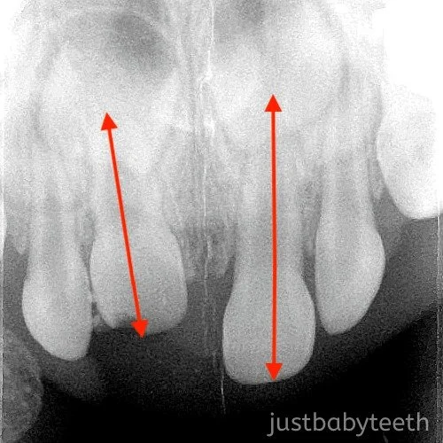
🔑: This case highlights to importance of radiographs in comprehensive exam. The patient presented with what appeared to be an avulsion. #E was completely absent in the clinical examination and the mom was unable to find the tooth. As difficult as it was to take a radiograph on a crying child, it’s not a step you want to skip. Based on the radiograph, you can see #E is still there, just way up there. Proper diagnosis is important to determine what the next steps are. The primary incisor’s root can go one of two ways: palatally (towards the primary tooth bud) or labially (away from the primary tooth bud). Based on the radiograph, you can see #E is foreshortened (compared to #F), which means the root tip is luxated labially. That’s good news and means that monitoring for spontaneous repositioning is an option.
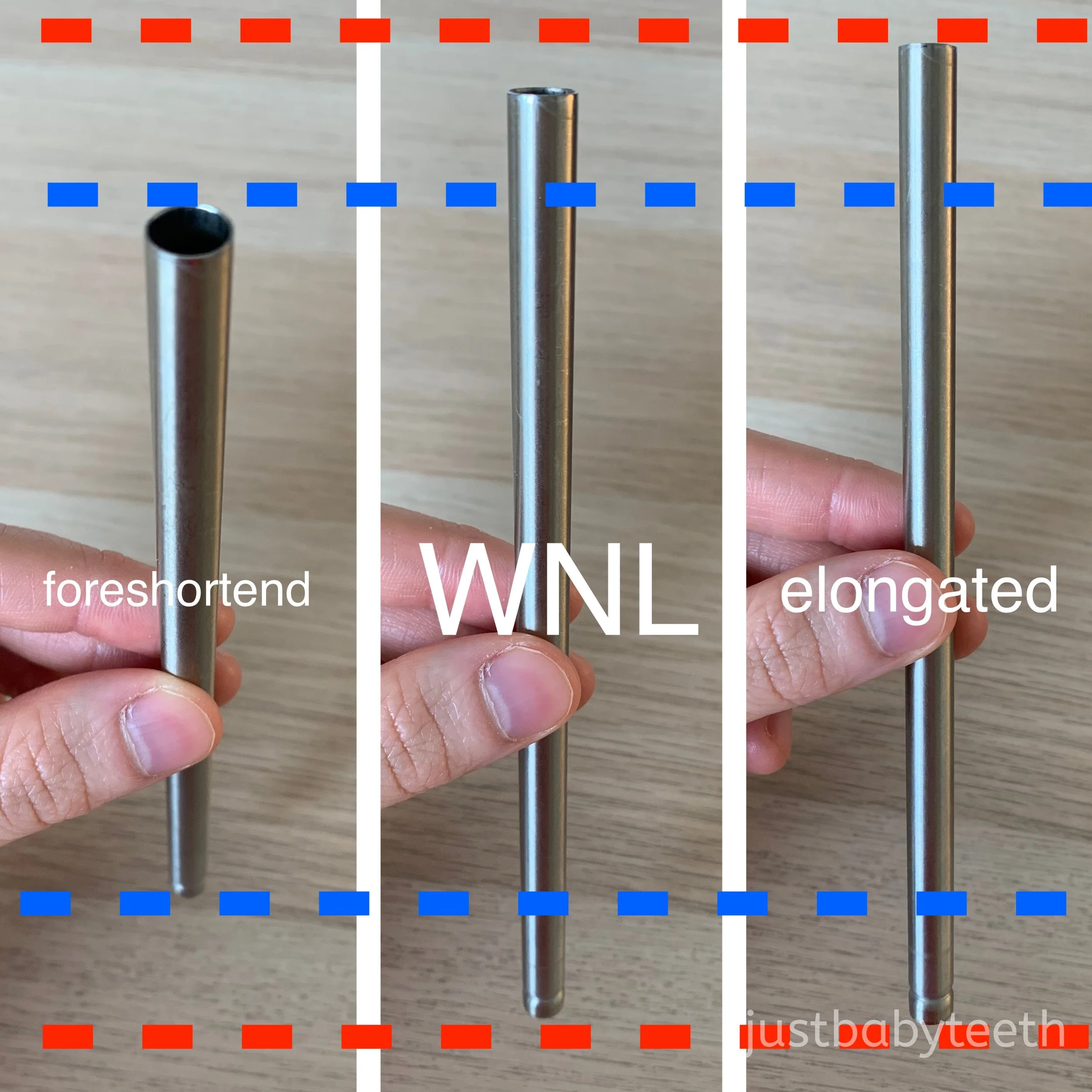
Wrapping your head around why foreshortening equals labial luxation and elongation equals palatal luxation can be difficult. I know it was for me! The third picture shows a straw taken slightly from above (as you were take an occlusal film). In the middle is the “normal position”. On the left, the straw is rotated towards the camera (labially) and appears shorter (foreshortened). On the right, the straw is rotated away the camera (palatally) and appears longer (elongated). I hope that helps!